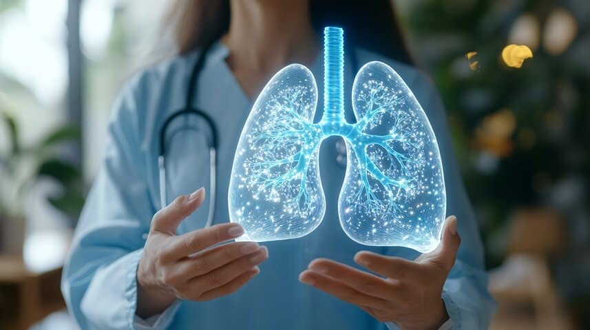
Understanding resilience – the ability of injured lung tissue to heal and regenerate – may hold the key to improving treatment and prevention of life-threatening lung diseases in extremely premature infants, according to a new study. Extremely preterm infants are those born at less than 28 weeks’ gestation. Using a four-dimensional microscopy technique, researchers at Vanderbilt University and Vanderbilt University Medical Center created 3D video images of lab-grown lung tissue from mice. What they found is nothing short of groundbreaking.
Premature Babies Often Have Impaired Lungs
“For the first time, we were able to image the forming lung live and quantify and measure the cell movements that together form an organ with sufficient surface area for gas exchange,” said Jennifer Sucre, MD, associate professor of pediatrics and cell and developmental biology. The group’s findings, published as the cover story in JCI Insight, the journal of the American Society of Clinical Investigation, represent a significant step toward improved treatment and prevention of bronchopulmonary dysplasia (BPD), which occurs in about 50% of infants born two to four months prematurely.

“If we understand how the lung forms, then we have a blueprint for how to grow new lungs after injury,” said the paper’s first author, Dr. Nick Negretti, a senior postdoc in Sucre’s lab who co-led the research. Mice have an extraordinary ability to repair the lungs, according to Sucre. Premature babies with BPD require oxygen and mechanical ventilation to breathe in the first few days after birth. However, oxygen therapy is a double-edged sword because it can also damage the delicate lung tissue. Although many premature babies can be weaned off the ventilator after a few days, they are at increased risk of developing serious breathing problems later in life, including chronic obstructive pulmonary disease.
Respiration – the exchange of oxygen for carbon dioxide – takes place in the alveoli via a delicate basement membrane between epithelial cells and blood vessels. According to the traditional view of lung development, invaginating septa (partitions) arise from a layer of epithelial, endothelial, and mesenchymal cells to subdivide the airspaces into alveoli.
However, when the researchers made and examined cross sections of the lungs of newborn mice over a period of three days, a different picture emerged: a balloon-like outgrowth of epithelial cells supported by a ring of myofibroblasts, i.e. cells that promote tissue formation. The innovative technology used in the Sucre laboratory enables the examination and identification of the specific molecules and signaling pathways that control this process. It also serves as a research tool for drugs that can promote tissue regeneration after injury.
Related Posts
-
Premature birth can affect the development of a child and seeking proper assessments from your…
-
When pregnant there are many concerns you might have about delivery and birth, one of…
-

When labor occurs prior to week 40 of gestation, delivery is categorized as a premature…